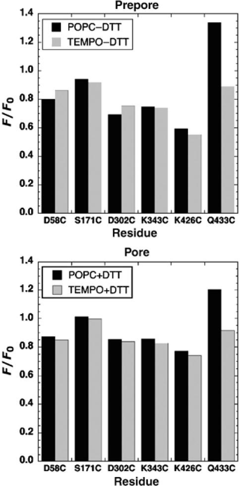
Figure 5.

Determination of residues in close proximity to the membrane surface in both prepore and pores by collisional quenching. The extent by which the fluorescence from selected labeled residues is reduced in bilayers that contain lipids with a quenching moiety in the headgroup region (TEMPO-PC), compared with the fluorescence from similarly modified proteins in bilayers without the TEMPO-PC (POPC), is shown. The residues denoted on the X-axis were mutated to cysteine and labeled with the fluorescent probe in the disulfide-locked mutant, PFOS190C-G57C. The extent of quenching was determined in both prepore and pore complexes of each dye-labeled mutant. Fluorophores located near the surface of the membrane surface will be quenched by the TEMPO-PC present in the liposomes. Legend: POPC−DTT, PFOS190C-G57C incubated with POPC-cholesterol liposomes as a prepore complex (disulfide remains oxidized); POPC+DTT, PFOS190C-G57C incubated with POPC-cholesterol liposomes as a pore complex (disulfide is reduced to allow prepore-to-pore conversion); TEMPO−DTT and TEMPO+DTT (same as POPC−DTT and POPC+DTT, except that 10% of the total lipid is replaced with TEMPO-labeled lipid). F/F0, ratio of fluorescence of membrane-bound PFO (F) to that for the soluble monomer (F0).
