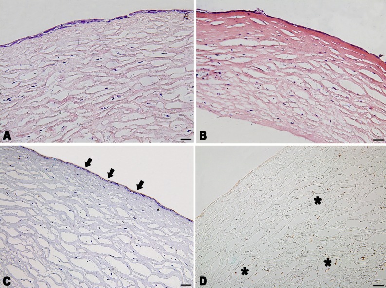
Fig 3. Phenotype and histological sections of the constructed rabbit anterior cornea replacement.
(A) H&E staining showed two or three layers of epithelial cells formed on the scaffold with a relatively uniform cell distribution in the matrix. (B) H&E staining of the construct soaked in 100% sterile glycerol for 5 minutes. (C) Immunohistochemistry staining for cytokeratin 3 (brown, black arrow) counterstained with hematoxylin (blue), and for vimentin (brown, asterisk) without hematoxylin staining. Scale bar: 50 μm.

