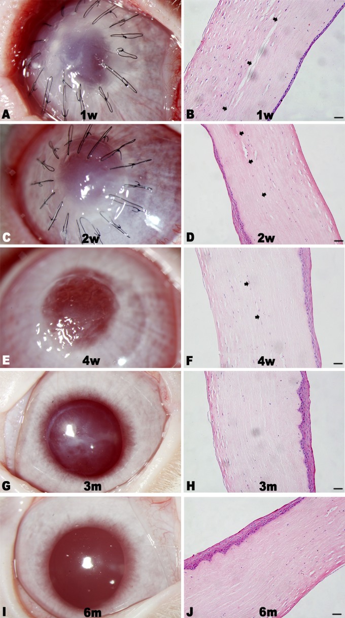
Fig 4. Representative images and histological sections of implanted corneas postoperative 6 months.
The implanted corneas gradually cleared with no apparent neovascularization (A, C, E, G, and I), and the constructs integrated well with the host cornea tissues with regular collagen arrangement as shown by H&E staining (B, D, F, H, and J). The boundary between the implant and host tissue was clear at the 4-week follow-up (B, D, and F, black arrows), and became indistinct 3 months (H) and 6 months (J) after surgery. Scale bar: 50 μm.

