
Figure 8.
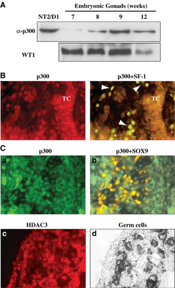
p300 and HDAC3 are expressed in the male embryonic gonad at the time of sexual differentiation. (A) Male human embryonic gonads at 7, 8, 9 and 12 weeks development stage and NT2/D1 as a control were lysed. Proteins (20 μg) were resolved by SDS–PAGE and blotted onto a nitrocellulose membrane. Immunoblots were performed using anti-p300 polyclonal (1 μg/ml) and anti-WT-1 (1/500) antibodies and were revealed using the ECL kit (Amersham). (B) An 8-week human male gonad section was co-immunostained with SF-1 antibody (de Santa Barbara et al, 1998) (in green) and p300 antibody (1/100, in red). At this stage, cells colabelled with both antibodies were identified as Leydig cells (indicated by an arrowhead), whereas Sertoli cells within the testicular cord (TC) were stained by p300 antibody only (magnification × 100). (C) p300 and HDAC3 proteins are expressed in 12.5 dpc male mouse gonads. p300 immunostaining (in green) was detected in most of the gonadal cells and colocalised with SOX9 (in red) in Sertoli cells. HDAC3 immunostaining (in red) was detected in the different gonadal cell types. Alkaline phosphatase staining of primordial germ cells (in black) localised gonadal tissue (magnification × 40).