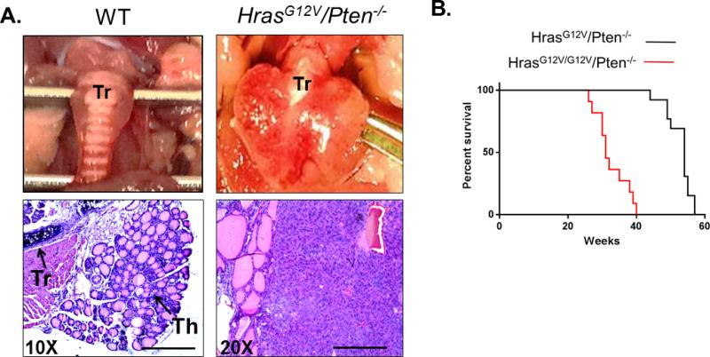
Figure 1.
HrasG12V and Pten loss leads to FTCs that progress to PDTC in HrasG12V/Pten−/−/TPO-Cre mice. (A) Top panel: Gross histology of wild-type (WT, left) and HrasG12V/Pten−/− (right) thyroid at 50 weeks of age. Tr=Trachea. HrasG12V/Pten−/− thyroid is significantly enlarged. (A) Bottom panel: H and E staining of 5 μm thick tissue sections from WT (left) and HrasG12V/Pten−/− (right) thyroid tissue. Th=Thyroid tissue, Tr=Trachea. WT thyroid contains organized follicles filled with colloid, in contrast to HrasG12V/Pten−/− thyroids, in which the normal follicular architecture is disrupted. 20X magnification, scale bar is 100 μM. (B) Survival of mice with heterozygous activation of HrasG12V (black line) is 100% at one year of age, while homozygous activation of HrasG12V (red line) is lethal by 40 weeks of age, with no mice surviving past this time point.

