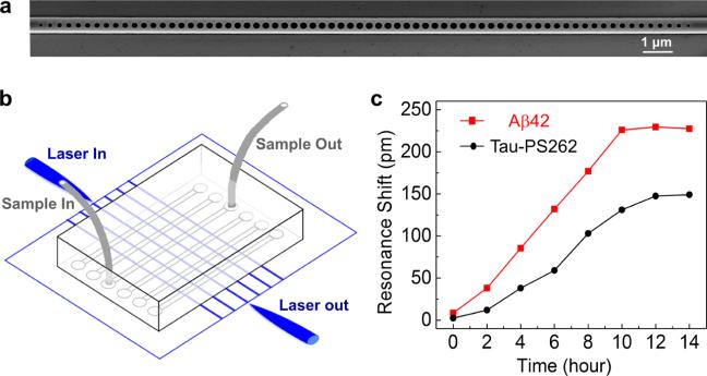
Figure 5.
Aβ42 and tau-PS262 detection in the SH-SY5Y-tau cell lysates using photonic crystal nanosensors. (a) Scanning electron microscope (SEM) image of the photonic crystal nanobeam cavity. (b) Schematics of the sensor chip, where light paths are denoted in blue and fluid paths are denoted in gray. The sensor chip is fabricated in silicon-on-insulator (SOI) platform. Telecom lasers were coupled to the edge of the chip and collected from the opposite side. Microfluidic channels were bonded on the chip for sample delivery. (c) Resonance shifts of the photonic crystal nanosensors measured in different cell lysates collected at different times after the isoflurane was applied.

