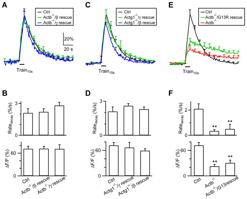
Figure 7. Actin polymerization is needed for endocytosis.
(A–B) FSypH traces (A), Ratedecay and ΔF (B) induced by Train10s (bar) in Ctrl hippocampal boutons (n = 8 experiments), in Actb−/− boutons transfected with β-actin (Actb−/−/β rescue; transfection of β-actin with SypH and a Cre-mCherry plasmid in ActbLoxP/LoxP boutons; n = 6), and in Actb−/− boutons transfected with γ-actin (Actb−/−/γ rescue, n = 6). Data are expressed as mean + s.e.m.
(C–D) Similar to A–B, but for Actg1−/− boutons transfected with γ-actin (Actg1−/−/γ rescue, n = 8) or β-actin (Actg1−/−/β rescue, n = 8).
(E–F) Similar to panel A–B, but for Ctrl boutons (n = 8), Actb−/− boutons (n = 11), and Actb−/− boutons transfected with β-actin(G13R) [Actb−/−/G13R rescue: transfection of β-actin(G13R) with SypH and a Cre-mCherry plasmid in ActbLoxP/LoxP hippocampal boutons; n = 8]. **: p < 0.01, t test.

