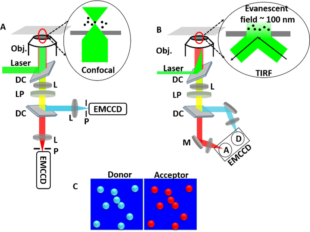
Fig. 3.
A. smFRET in confocal microscopy. Laser is reflected by a dichroic mirror and then focused to sample by an objective (Obj.) with a high numerical aperture (NA). The fluorescence signals from donors and acceptors are passed through a long-pass filter (LP) and then split by a dichroic (DC) into two different channels. Both signals are focused to two separate APDs by lens (L). Before APDs, there are two pinholes (P) to reduce fluorescence signal originated from above and below of the focal plane. Inset is showing the configuration of excitation laser on the sample. B. smFRET in objective type total internal reflection fluorescence microscopy (TIRF), which exploits the unique properties of an induced evanescent wave or field in a limited specimen region (~100 nm) immediately adjacent to the interface between two media having different refractive indices. Fluorescence signals from donors and acceptors are focused on same EMCCD. Inset shows the configuration of the laser on the sample. C. Donor and acceptor data are recorded simultaneously and shown in cyan and red respectively.

