
Fig. 1.
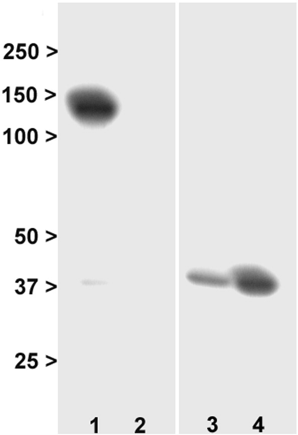
Characterisation of membrane and cytosolic fractions. Lane 1: membrane fraction, N-cadherin. Lane 2: cytosol fraction, N-cadherin. Lane 3: membrane fraction, GAPDH. Lane 4: cytosolic fraction, GAPDH. The blots show the predicted localisation of N-cadherin to the membrane, and the enrichment of GAPDH in cytosol. Numbers are molecular weight (kDa).