Figure 1.
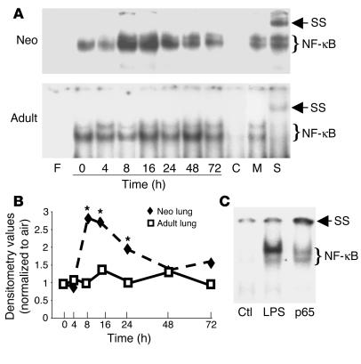
Demonstration of hyperoxia-mediated neonatal (Neo) lung NF-κB activation in C57BL/6 mice. (A) Representative of 3 EMSA blots. F, free probe; 0–72 h, duration of hyperoxia; C, competition with 100-fold excess of unlabeled oligonucleotide probe; M, competition with 100-fold excess of unlabeled mutated oligonucleotide probe; S, supershift with p50 antibody in lung extract collected at 8 hours of hyperoxia; SS, supershift retardation band. (B) Densitometric evaluation of the EMSA blots. Densitometric units were expressed as a ratio of air-exposed values. The data are the mean ± SE of 3 separate experiments. *P < 0.05 vs. similarly exposed adults. (C) NF-κB activation in the neonatal LPS-injected lung. LPS (20 mg/kg) was injected intraperitoneally into 5-day-old C57BL/6 mice. Ctl, mice injected with PBS as vehicle control; LPS, 1 hour after injection with LPS; p65, sample as in LPS lane incubated with p65 antibody. Note: The decreased NF-κB complex and the supershift gel retardation band demonstrating the binding complex are specific to p65 in this model.

