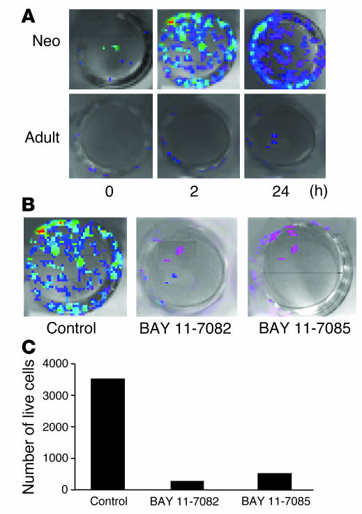
Figure 7.
Effect of inhibition of I-κΒα phosphorylation on primary lung cells exposed to hyperoxia. (A) Visualization of NF-κB activation in primary lung cells cultured from NF-κB/luc Tg mice. (B) Effect of inhibition of I-κΒα phosphorylation on NF-κB activation. Cells were incubated with 1 mM BAY 11-7085 or BAY 11-7082 and then exposed to 24 hours of hyperoxia. Controls were incubated with 0.1% ethanol, the vehicle for BAY. Note the decreased light intensity after BAY treatment. (C) Cell viability was evaluated using trypan blue exclusion. The number of surviving cells in each group was assessed after 24 hours of hyperoxic exposure.

