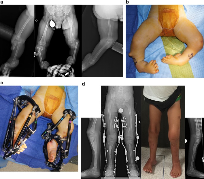
Fig. 10.
a Preoperative radiographs of a 13-month-old boy with bilateral tibial hemimelia; Paley type 4A tibial hemimelia on the right and Paley type 4B tibial hemimelia on the left. Both sides have severe equino-varus deformities of both feet. The proximal tibial remnant on the right is prominent under the skin. There is proximal migration of the fibula on both sides. There is an unossified proximal tibial cartilagenous anlage on the left side. b Clinical photograph showing the severe equino-varus feet. Note that the tip of the proximal tibial remnant has its own skin pouch. c Bilateral TSF devices were applied to both legs. The thigh rings are perpendicular to the femurs and the foot rings are parallel to the soles of both feet. This computer-dependent external fixator is programmed first to correct the deformities between the femur and fibula at the knee, including transporting the fibula distally. It is then reprogrammed to correct the foot deformity. No additional surgery is required to switch from knee to ankle correction. On the left leg the proximal fibular epiphysis was transported distally and brought under the tibial epiphysis. d Standing AP photograph (center right) and radiograph (center left) of both lower limbs at age 5 years, showing that the legs are well aligned. Lateral radiographs of right leg (left) and left leg (right) showing the feet are fused at the ankle in a plantigrade position. Both distal fibular physes are closed despite best efforts to preserve them. The right foot has some adductus supination deformity. The patient is shown after lengthening of both lower limbs was completed. The left proximal tibial epiphysis is ossified and fused to the proximal fibular epiphysis. The unossified proximal tibial epiphysis was ossified by insertion of BMP2 into drill holes in the cartilage of the tibial anlage. The ossification of the tibial epiphysis and fusion to the fibula occurred within 3 months. Note, there is preservation of the proximal physis of the fibula. On the right the fibula was transferred to the tibia. It has auto-bridged across to the proximal fibula. Initially, knee–ankle–foot–orthotic (KAFO) braces were used. The knee was stable enough to discontinue these after these radiographs

