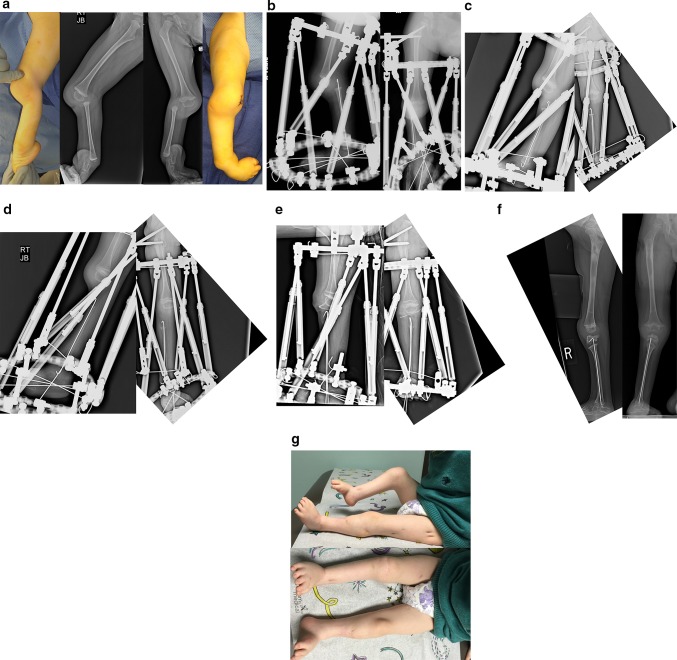
Fig. 11.
a Lateral (left and center left) and AP (right and center right) photographs and radiographs of the right leg of a 2-year-old boy with bilateral tibial hemimelia. Only the right side is shown. The right side has Paley type 5A tibial hemimelia and the left side Paley type 4A. Note the flexion contracture of the right knee and the equino-varus-adductus foot deformity. b Lateral (left) and AP (right) radiographs showing the TSF in place with femoral and foot rings. The fibula is secured to the foot ring with a transverse wire. Note the temporary epiphysiodesis wire in the fibula hooked at both ends. Also note the wire across the neck of the talus. c Lateral (left) and AP (right) radiographs at the end of gradual distraction with TSF. The fibular head is centered under the end of the femur. The foot position has not changed and the transverse fibular wires remains connected to the foot ring. d Lateral (left) and AP (right) radiographs showing that the transverse fibular wire was fixed to the proximal ring using a threaded rod and post. This wire was released from the distal ring to allow gradual correction of the equino-varus foot contracture and to bring the talus under the distal fibula. A second computer planning is carried out to generate a new adjustment schedule for the patient. Note that there is no ossification of the proximal fibular epiphysis. Also note the growth lines related to preoperative infusion of zolidronic acid to prevent disuse osteoporosis during distraction. e Intraoperative lateral (left) and AP (right) radiographs after the surgery to perform a patellar arthroplasty and fibula–talar fusion is completed. Note the hemovac drain can be seen at the knee. The Paley–Weber patellar arthroplasty was performed fixing the proximal fibular epiphysis to the patella with a hooked intramedullary wire secured to the bottom of the frame below the foot. The talus was fused to the distal tibial epiphysis. BMP2 was inserted into a drill hole in the patella and proximal fibula to lead to ossification and fusion of both the patella and fibular epiphysis. A transverse distal fibular wire is arced to the foot ring to apply compression across the fusion site. f Lateral (left) and AP (right) radiographs showing the patella and proximal fibular epiphysis are ossified and fused together with preservation of the proximal fibula physis. The patella now serves as a tibial plateau. The talus has also fused to the distal fibula but the distal fibular physis has closed. g Clinical photographs, frontal view (bottom) and medial view (top) showing that the right knee can bend to 90° (top). There is active motion present. The other leg was also successfully treated for type 4A tibial hemimelia

