Fig. 12.
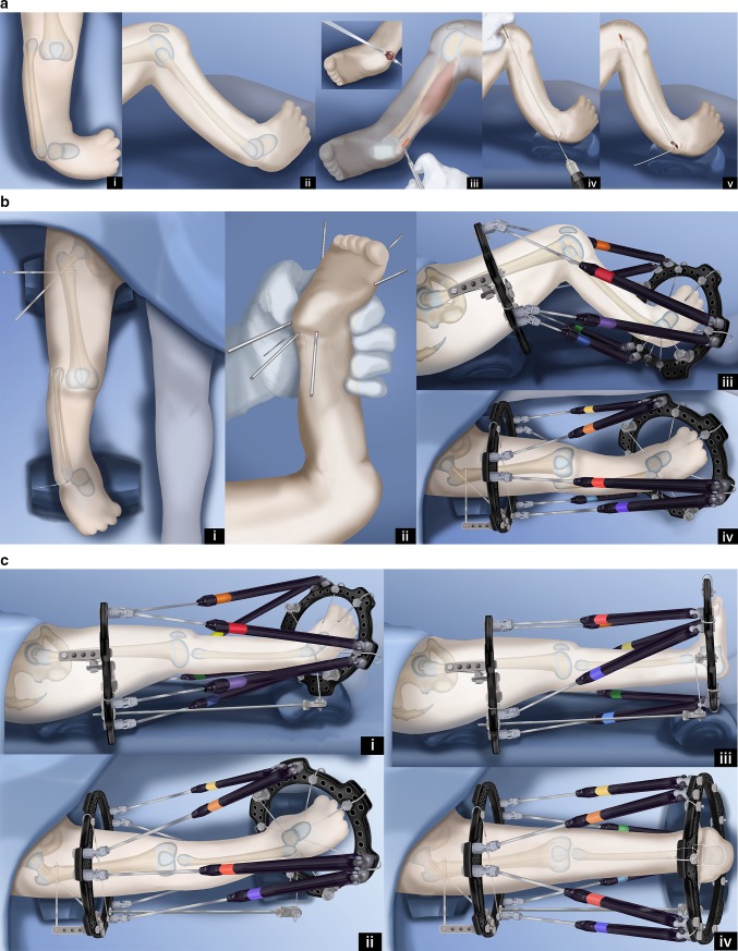
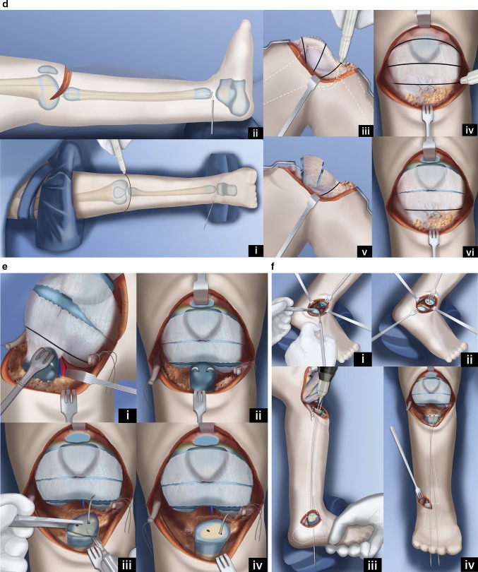
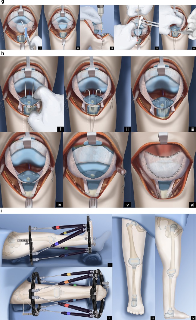
Paley–Weber patellar arthroplasty. a Frontal (i) and lateral (ii) view illustrations of Paley type 5A tibial hemimelia: complete tibial agenesis, patella present, fibula dislocated and proximally migrated, knee flexion contracture and fixed equino-varus foot deformity. First surgery consists of Achilles tenotomy (iii). Two 1.5-mm intramedullary wires are inserted retrograde into the fibula and both are curled and hooked into the proximal fibular epiphysis. One is curled around the distal fibular epiphysis and the other is bent 90° to stick out of the skin (iv, v). Reproduced with permission by the Paley Foundation. b A prophylactic wire is inserted in the femur and hooked over the greater trochanter. Two proximal femoral half pins are inserted—one at the level of the lesser trochanter and one up the femoral neck (i). Three wires are inserted in the calcaneus in the plane of the sole of the foot—one mid posterior to exit near the interspace of the first and second toes; one posterolateral to exit anteromedial; one posteromedial to exit anterolateral (ii). The proximal femoral ring is fixed to the two half pins and an olive wire is added from antero-medial to postero-lateral in the mid femur. A foot ring is fixed to the three wires in the foot and a transverse talar wire added. The 90° bent fibular wire is fixed to the foot ring. Finally, six struts are added between the rings. Computer planning is done to gradually correct the knee deformity (iii, iv). Reproduced with permission by the Paley Foundation. c After several weeks of gradual correction of the knee joint contracture, the fibula is centered on the femur. At this point the fibular wire is connected to a long threaded rod with post and then liberated from the foot ring (i, ii). A new computer planning is carried out to gradually correct the foot deformity. The talus is repositioned under the end of the fibula. The knee and ankle are now ready for the next stage surgery (iii, iv). Reproduced with permission by the Paley Foundation. d Second surgery starts with removal of the foot ring and wires. The leg and proximal ring are prepped and draped, and the proximal ring is covered with a sterile towel. An Esmarch is used as a tourniquet. A transverse concave proximal incision is made at the level of the knee joint (i, ii) and the knee joint capsule exposed. Two “visor” flaps should be outlined with three lines that should converge near the lateral and medial aspects of the knee. The upper line passes across the top end of the patella and the middle line at the lower end of the patella. The most distal line passes as posteriorly as possible (iii, iv). The superior pole of the patella is cut through as part of the upper incision, and a second incision is then made below the lower end of the patella (v, vi). Reproduced with permission by the Paley Foundation. e Before the distal part of the lower visor flap is incised, the biceps tendon is detached laterally and the semitendinosis tendon medially. The lateral border of the medial head of the gastrocnemius is located, and the popliteal vessels are identified (i). Staying clear of the vessels cut the posterior limb of the inferior visor flap (ii). The hook in the fibular wires is unfolded and one wire is removed. The epiphysis of the proximal fibula is cut through to expose its ossific nucleus (iii, iv). Reproduced with permission by the Paley Foundation. f A transverse incision is made at the lateral aspect of the tip of the lateral malleolus and the distal fibula and talus exposed. One fibular wire is removed and the second uncurled. The talus is cut across parallel to the sole of the foot and the distal fibula is cut across perpendicular to the fibula, exposing the bone of both ossific nuclei in preparation for fibula-talar fusion (i, ii). Remove the remaining fibular wire and replace the previous wires with two new wires. These wires are advanced through the fibula and drilled across into the talus and out the plantar aspect of the foot to fuse the fibula to the talus (iii, iv). Reproduced with permission by the Paley Foundation. g Slide the proximal visor flap which contains the patella posteriorly, underneath the distal visor flap, which moves anteriorly (i). The anterior surface of the cartilagenous patella is exposed by reflecting back two perichondral flaps (H flap; ii). A small hole is drilled in the patella from proximal to distal and a second hole is drilled to intersect the first in a T-junction from anterior towards posterior (iii). The articular surface must not be penetrated. A small shallow hole is also drilled in the epiphysis of the fibula but not deep enough to reach the physis. A BMP2 sponge (INFUSE® Bone Graft; Medtronic, Dublin, Ireland) is inserted into the holes to induce ossification of the patella and patella–fibular fusion (iv). The anterior and posterior holes in the patella are plugged with bone wax to prevent leakage of the BMP2 (v). Reproduced with permission by the Paley Foundation. h The retrograde fibular wires are advanced through the patella (i), then bent over themselves and the hook pulled back into the patella (ii). The apex of the bend must be submerged in the cartilage of the patella (iii). The perichondral flaps are sutured to the medial and lateral sides of the fibula (iv). The patella now sits conformed and congruent with the distal femur to act as a tibial plateau (v). The visor flaps are sutured together. The quadriceps muscle and patellar remnant are sutured to the proximal edge of the superior visor flap, and the the biceps and semitendinosis tendons are sutured to the lateral and medial aspects of the fibula, respectively (vi). Reproduced with permission by the Paley Foundation. i After the incision is closed layer by layer, three wires are inserted in the foot, a foot ring applied and six struts connected. One transverse distal fibular wire is arched and tensioned to compress the ankle fusion. The axial wires are fixed below to the under surface of the distal ring. The foot is plantigrade and the knee is well centered and aligned. The frame stays on the leg another 3–4 months (i, ii). After the external fixator is removed the hook wires are left in place and cut short to stay buried in the calcaneus (iii, iv). Reproduced with permission by the Paley Foundation


