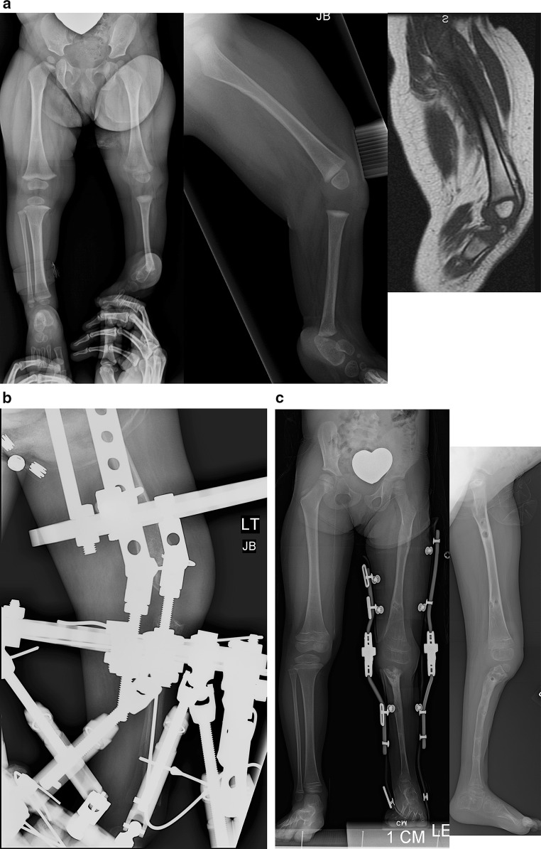
Fig. 13.
a Antero-posterior (left) and lateral (middle) radiographs of a 12-month-old girl with Paley type 5B unilateral tibial hemimelia. The fibula is hypertrophied and centered on the femur. The foot is in equinovarus. A sagittal section of the magnetic resonance imaging scan (right) shows an absent patella, with a quadriceps muscle extending to the fibula. b Lateral radiograph showing a TSF device with six struts applied between the upper fibula and foot to correct the equino-varus foot deformity and to bring the talus under the end of the fibula. An Ilizarov apparatus on the femur connects to the TSF with hinges at the knee joint. A distraction mechanism is in place to correct the knee contracture while permitting removal of this mechanism to allow physical therapy to move and exercise the knee through the hinges. c Standing AP (left) and lateral (right) radiographs showing the fibula is centralized, hypertrophied and lengthened. A KAFO brace is used to protect the stability of the knee and promote hypertrophy. The talus is fused to the fibula with the foot in a plantigrade position. The distal and proximal fibular physes are both patent

