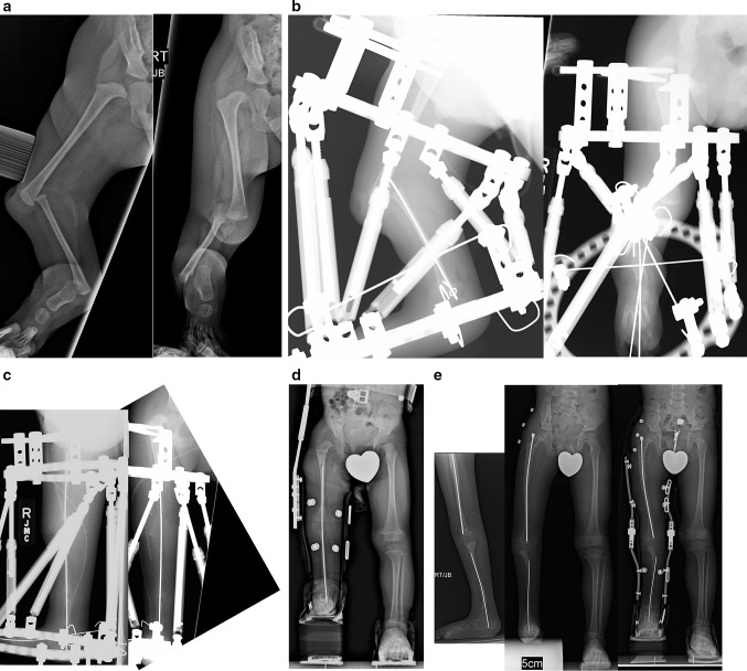
Fig. 14.
a Preoperative AP (left) and lateral (right) radiographs of the right leg of a 15-month-old boy born with unilateral tibial hemimelia, Paley type 5C. There is no patella or quadriceps muscle. The knee and ankle have severe contractures. b Lateral (left) and AP (right) radiographs showing TSF in place with one ring on femur and one on the foot. There is a temporary epiphysiodesis wire in the fibula hooked at both ends. There is a transverse wire through the fibula to transport it distally. c Intraoperative (hemovac seen) lateral (left) and AP (right) radiographs after complete correction of the knee and ankle and after the second staged surgery to fuse the ankle and to temporarily arthrodese the knee using an axial wire. Tendon transfers were also done to replace the absent quadriceps muscle. d AP standing radiograph following removal of external fixator 3 months later. The axial wire was left in place to protect the knee for 6 more months. There is excellent alignment with significant leg length difference. e Standing lateral (left) and AP (middle, right) radiographs at age 5 yers, after lengthening of the femur and tibia and after a pelvic osteotomy was done to stabilize the hip. Separate intramedullary wires remain in place to protect the bones from fracture and to guide hypertrophy. A KAFO is used for several years—until the knee becomes stable—to allow knee flexion and extension while protecting varus and valgus bending on the hypertrophying knee joint for several years until the knee becomes stable

