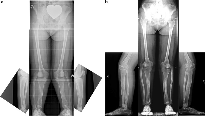
Fig. 4.
a Antero-posterior (AP) and lateral radiographs of 20-year-old woman with bilateral Paley type 1 tibial hemimelia. The tibia is well formed at both the knee and ankle joints. The fibulas are relatively overgrown at their proximal ends and are articulating with the side of the femurs. The knees are both in valgus due to both tibial and femoral deformities. The tibias have mild procurvatum diaphyseal bowing. Since both tibias are relatively short compared to the femurs, the patient has a mesomelic disproportion and short stature. b AP and lateral radiographs after treatment. Both tibias were lengthened with external fixators. The tibial valgus-procurvatum was corrected. The fibulas were not pulled down. Bilateral femur varus osteotomies were performed after completion of the tibial correction. The lengthening restored the proportion of the tibias and femurs to normal

