Fig. 5.
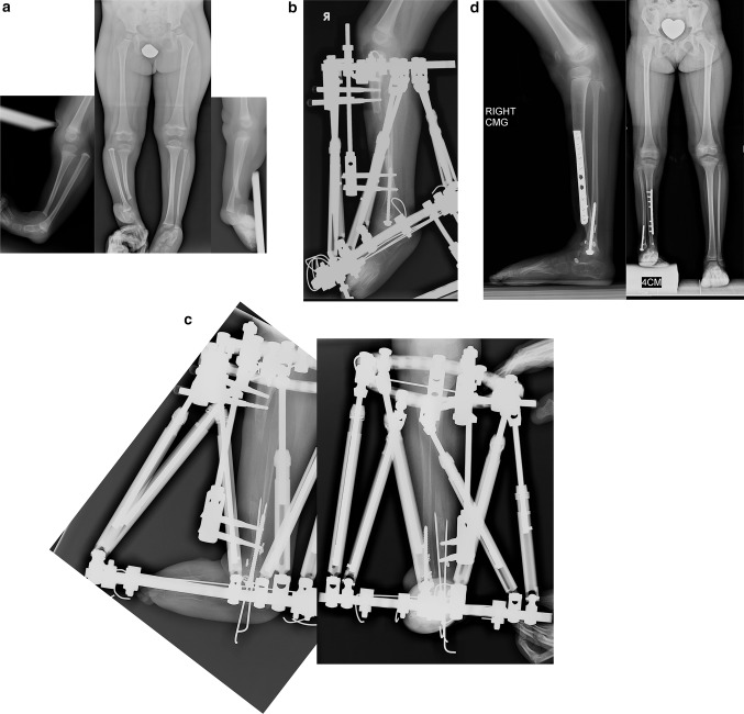
a Radiographs of 2-year-old girl with Paley type 2A unilateral tibial hemimelia, with equino-varus deformity of foot and varus of tibial diaphysis. There is no diastasis of the distal tibio-fibular joint. The ankle joint is present. The ossification of the distal tibia shows a regular trumpet shaped metaphyseal line indicating that this is the region of the distal tibial physis. b Lateral radiograph of tibia. Taylor spatial frame (TSF) is in place with proximal tibial and foot rings programmed for gradual correction of foot deformity. There is also an independent mechanism (threaded rod with cube and two half pins connected to the proximal ring) to simultaneously lengthen the tibia through a proximal osteotomy. There is a distal fibular epiphysiodesis screw in place to slow the growth of the faster growing fibula. c Lateral (left) and AP (right) radiographs at the end of the foot correction and 8 cm of lengthening after a second stage surgery to openly reduce the talus to the tibia combined with axial pinning of the ankle joint. Syndesmotic washer-suture device (Ziptite™ Fixation System; Biomet Sports Medicine, LLC, Warsaw, IN) inserted to stabilize the lengthened tibia to the fibula at its new syndesmosis level. The tibial lengthening bone shows poor regenerate formation. d Plate fixation at time of removal of fixator to prevent fracture. The tibia is well aligned. The talus is under the distal tibia. The fibula is now at station at the ankle and the ankle is stable. The foot is plantigrade

