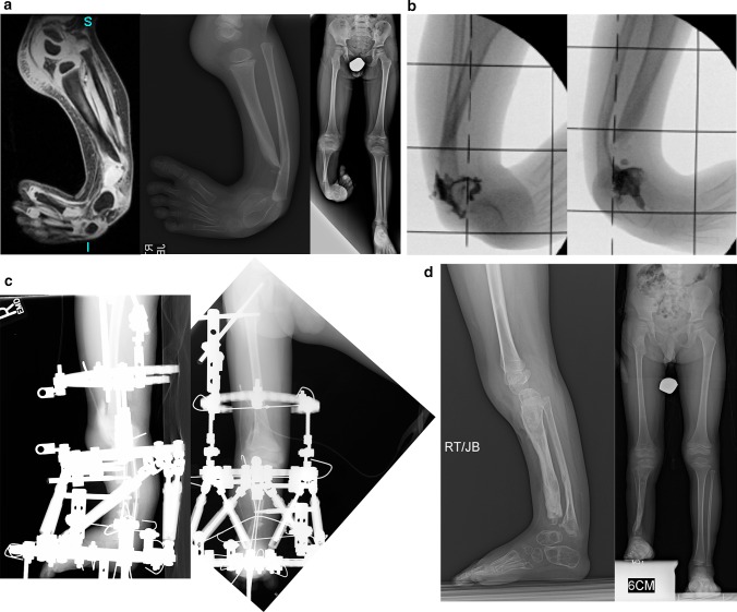
Fig. 7.
a Magnetic resonance imaging (MRI) scan (left), lateral radiograph (middle), and standing AP radiograph (right) showing Paley type 2C unilateral tibial hemimelia. The foot is in significant varus. The distal tibia has an unossified region contiguous with the ankle. There is no obvious distal tibial physis, but the plafond is present. Note that the ossification of the distal tibia is irregular and sloped, not like a normal metaphysis associated with a distal tibial physis (Fig. 5a). The proximal fibula is relatively overgrown and proximally migrated. b Arthrogram of ankle showing AP (left) and lateral (right) views. c Lateral (left) and AP (right) radiographs after arthrotomy of ankle, and open valgus-extension osteotomy and shortening of distal tibia to realign ankle joint by acute correction. Bone morphogenetic protein 2 (BMP2) was inserted in drill holes of the cartilage remnant of the distal tibia to get it to ossify. A second osteotomy was performed proximally for lengthening. The fibula is being pulled distally with an intramedullary wire hooked over the proximal epiphysis (see AP view). Because the tibial/ankle/foot deformities were corrected acutely, the external fixator is programmed for pure lengthening. The external fixator is extended to the femur with knee hinges to protect the cruciate deficient knee during lengthening while permitting knee flexion and extension motion. d Lateral (left) and AP (right) radiographs, after lengthening with excellent consolidation of the tibia, including ossification of the delayed distal tibia. The foot is plantigrade with forefoot supination deformity present. The proximal fibula is at station

