Fig. 8.
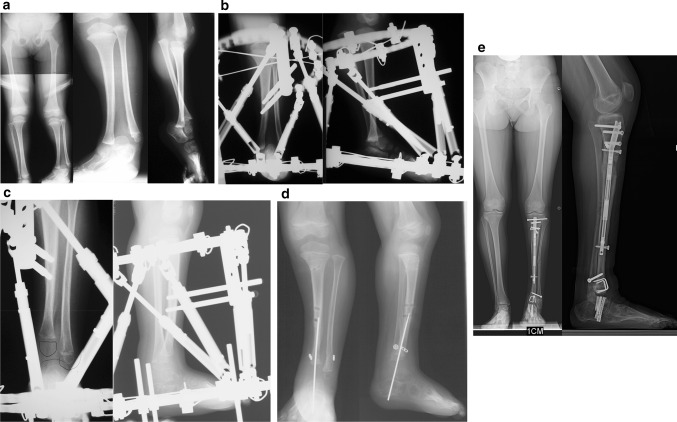
a Standing (left), mortis view (middle) and lateral view (right) radiographs of a 2-year-old girl with Paley type 3A unilateral tibial hemimelia. The diastasis of the distal tibia and fibula with the talus in between is very evident. The talus always remains in line with the distal fibula and together they internally rotate around the distal tibia. The talus is shortened relative to the distal tibia but not relative to the distal fibula. There is relative overgrowth with proximal migration of the fibula. There is a mild distal tibial varus diaphyseal bowing. b AP (left) and lateral (right) radiographs showing TSF applied to the tibia and foot. There is no fixation in the fibula or talus. More current constructs would fix to these as described in the text. The foot is in internal rotation and equinus. c AP (left) and lateral (right) radiographs after gradual correction using TSF. The talus is under the distal tibial epiphysis and the foot and fibula have rotated externally relative to the tibia. There is no longer any tibio-fibular diastasis. The equinus deformity is also corrected. This gradual correction took 12 weeks. d AP (left) and lateral (right) radiographs after removal of the external fixator 3 months following open ankle reconstruction. The ankle reconstruction included a biologic arthroplasty to create a concave surface on the distal tibial epiphysis that is congruent to the talar dome convexity. The ankle joint was pinned with an axial wire and the diastasis of the tibia and fibula were fixed with a suture-washer syndesmotic repair (TightRope®, Arthrex, Naples, FL). The axial wire was left in place for another 3 months to prevent recurrent deformity at the ankle. Note the proximal fibula was brought down to station. e AP (left) and lateral (right) radiographs at age 14 years after she underwent two successful lengthenings; one external fixator lengthening at age 6, and one implantable nail lengthening at age 14 (Precice™; NuVasive Inc., San Diego, CA). She also had a supramalleolar and subtalar osteotomy. Note the ankle joint is stable and well preserved. The ankle has about 20° of motion. The proximal fibula is at station. Most of this hardware was removed at a later date. Due to the limited ankle motion, the leg lengths were intentionally corrected to leave a 1-cm difference

