Fig. 9.
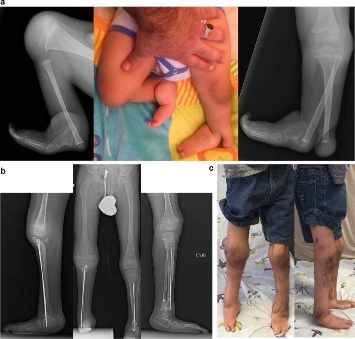
a Preoperative photograph (center), and radiographs (right on the left, left on the right) of bilateral tibial hemimelia in a 3-year-old boy; Paley type 5A (right) and Paley type 3B tibial hemimelia (left). The patella on the right has not yet ossified. Other than the cleft on the left, this is very similar to type 3A. The cleft makes this a very rare type. It should be noted that the foot is found on the fibular side of the cleft on the left. Both feet are in severe equino-varus and even upside down. There is a greater than 90° flexion contracture and dislocation at the right knee. b Standing (center), right lateral (left) and left lateral (right) radiographs after the staged reconstruction method was completed, 2 years prior on the left and 3 months prior on the right. The Paley–Weber patellar arthroplasty was performed on the right, with insertion of BMP2 to ossify the patella and fuse it to the proximal fibular epiphysis. Note the preservation of the proximal fibular physis. On the left the talus was brought under the tibia, a biologic arthroplasty performed together with plastic closure of the cleft. The distal and proximal tibial and fibular physes are preserved on both sides. The fusion of the talus to the distal fibular epiphysis did not occur and may require revision in the future. Both feet are plantigrade. c Clinical photographs showing the patient standing with both feet plantigrade and both knees straight and well aligned

