Figure 4.
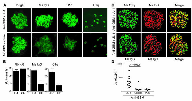
Effects of administration of anti-C1q mAb to mice pretreated with C1q-fixing anti-GBM antibodies. (A) Immunofluorescence of renal sections obtained from mice pretreated with rabbit (Rb) anti-GBM antibodies combined with either mAb JL-1 or IgG2b control mAb stained for the presence of mouse (Ms) IgG, mouse C1q, or rabbit IgG. Images show linear, GBM-like deposition of anti-GBM antibodies, linear fixation of C1q and anti-C1q in the JL-1–coinjected mice, and only mild mesangial positivity for C1q and anti-C1q in control-coinjected mice. Original magnification, ×400. Right panels: Low-power magnifications of renal sections of mice injected with anti-GBM in combination with either JL-1 or control and stained for C1q. Original magnification, ×100. (B) Quantification of immunofluorescence analysis of the glomerular deposition of rabbit IgG, mouse IgG, and C1q. Ctr, control. (C) Confocal analysis of kidney sections of mice injected with rabbit anti-GBM and JL-1. Representative pictures are shown for the colocalization (yellow) of mouse C1q (green) and mouse IgG (red) and the colocalization (yellow) of rabbit IgG (green) and mouse IgG (red). They indicate that rabbit IgG, mouse C1q, and mouse IgG do colocalize in these mice, in a linear, GBM-like pattern. (D) Albuminuria of mice injected with anti-GBM antibodies followed by JL-1, control mAb, or PBS.

