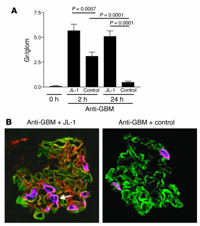
Figure 7.
Quantification of glomerular granulocyte influx. (A) Mice were injected with anti-GBM antibodies in combination with either JL-1 or control mAb. Granulocytes per glomerular cross section were scored either at 2 hours or at 24 hours after injection. (B) Confocal analysis of sections stained for mouse C1q (green), mouse IgG (red), and mouse granulocytes (purple). The pictures are merged and show, in yellow, colocalization of green and red, and, in white, colocalization of green and purple. The anti-GBM antibodies induced linear fixation of C1q in both groups, but only in the JL-1–coinjected mice is there colocalization between C1q and IgG and a pronounced influx of granulocytes. Original magnification, ×400. The white arrow indicates the white colocalization between C1q and granulocytes.

