Figure 2.
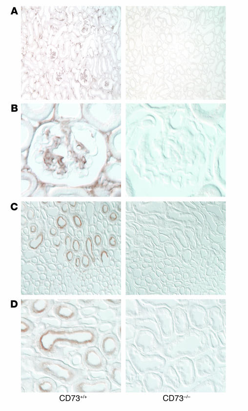
e-5′NT/CD73 Ab staining of kidney sections from WT and e-5′NT/CD73–deficient mice. (A and B) Immunoperoxidase staining of sections of the renal cortex with a polyclonal Ab against e-5′NT/CD73. In sections from WT mice (left) positivity was noted predominantly in the glomerular tuft, including the glomerular stalk, and in interstitial cells. Staining was absent in sections from e-5′NT/CD73–/– mice. (C and D) In the renal outer medulla e-5′NT/CD73 positivity was found in the brush border of proximal straight tubules (left), with no staining being detectable in sections from e-5′NT/CD73–/– animals. Magnification in A and C: ×100; in B and D: ×600.

