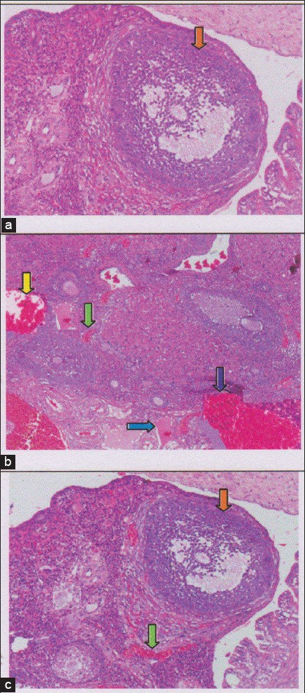
Figure-4.

The regeneration of ovarian tissue indicated by microscopic examination with hematoxylin and eosin (HE) staining in rat ovarian tissue in a few treatments. (a) Control negative group (T0−), with normal ovary without honeybee product was shown growing follicles ( ); (b) the group of ovary failure (T0+), congestion of ovarian (
 ), and severe hemorrhage (
 ), also visible hemosiderin (
 ) due to blood cell lysis (brownish yellow color) with deposition of fibrin (
 ) indicating that chronic congestive has been occurred, (c) the group ovary failure + 50% honeybee product (T1), ovaries begin to regenerate so it looks intact, although there is still a slight hemorrhage and congestion in some areas, but has seen growing follicles (
 ).
).
