Figure 1.
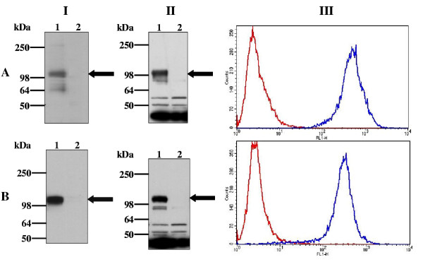
Expression of HA-S1P4-Gαi1 and HA-S1P4(E122Q)-Gαi1(C351I) in CHO-K1 cells. Membranes from untransfected CHO-K1 cells (lane 2) and CHO-K1 cells stably expressing HA-S1P4-Gαi1(C351I) (A) or HA-S1P4(E122Q)-Gαi1(C351I) (B) were analysed by Western blotting using anti-HA (panel I) or anti-Gαi1 (panel II) antibodies. Visualisation of immunoreactive proteins was achieved using chemiluminescence after incubation of the blot with appropriate HRP-conjugated secondary antibodies. The position of each HA-S1P4 fusion protein is indicated by an arrow. Cell-surface expressed HA-S1P4 receptor was detected by FACS analysis (panel III) using a Fluorescein conjugate of the anti-HA antibody (blue line). Cells were also stained with an isotype matched control antibody (red line). No staining of untransfected CHO-K1 cells was observed using the Fluorescein conjugate of the anti-HA antibody (not shown). Data are presented as overlay histograms and are representative of at least five independent experiments.

