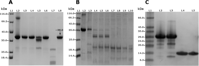
Figure 5. 12% SDS PAGE showing recombinant PCP protein autoproteolytic assays.
Figure 5 (A) Lane 1: Protein maker. Lane 2: Purified denatured protein at 95 °C as native control. Lane 3: Protein incubated at −21 °C showing no autoproteolysis. Lane 4: Protein incubated at −21 °C with protease inhibitors cocktail. Lane 5: Protein incubated at 4 °C showing partial autoproteolysis. Lane 6: Protein incubated at 4 °C with protease inhibitors cocktail with no autoproteolysis. Lane 7: Protein incubated at 25 °C showing complete autoproteolysis. Lane 8: Protein incubated at 25 °C with protease inhibitors cocktail showing that the autoproteolysis was inhibited. (B) Lane 1: Protein Marker. Lane 2: Purified denatured protein at 95 °C. Lane 3: through Lane 10: Purified protein incubated at 25 °C in different buffered pHs: 4.1, 4.8, 5.6, 6.1, 6.8, 7.2, 7.9 and 8.5, respectively. An increase in the autoproteolytic activity of PCP can be correlated with the increase in pH. (C) Lane 1: Protein marker; Lane 2: Purified construct of domains 1 through 3 (D13) incubated at 4 °C showing autoproteolytic activity; Lane 3: Purified D13 incubated at 25 °C and displaying autoproteolytic activity; Lane 4: Purified construct of domains 3 and 4 (D34) incubated at 4 °C and displaying no autoproteolysis; Lane 5: Purified D34 incubated at 25 °C and displaying no autoproteolysis.

