
Figure 3. Comparison of the activities of BA transporters between hiHeps and human hepatocytes.
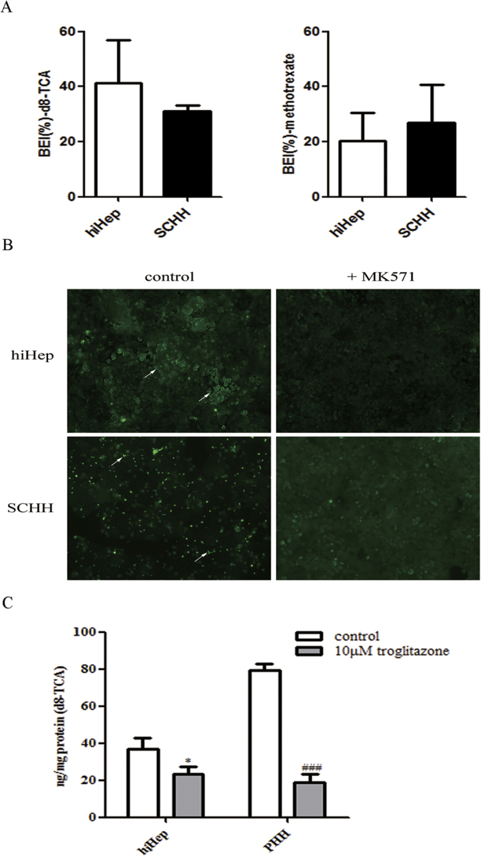
(A) The efflux transporter activities of BSEP and MRP2 were determined by calculating the BEI values of d8-TCA (the substrate of BSEP) and methotrexate (the substrate of MRP2) for hiHeps (white columns) and SCHHs (black columns). The data are expressed as the mean ± SD (n = 3). (B) The polarized locations of efflux transporters in the bile canaliculi of both hiHeps and SCHHs, as determined by fluorescence microscopy. In the absence or presence of MK571 (20 μmol/L), an MRP2 inhibitor, bile canaliculi were labelled with an MRP2 fluorescent substrate, CDF, which was formed from non-fluorescent CDFDA via intracellular esterases. (C) The influx transporter activity of NTCP was determined by accumulation assay of an NTCP substrate, d8-TCA. Troglitazone (10 μmol/L) was used as a positive control. The data are expressed as the mean ± SD (n = 3). *p < 0.05 vs control in hiHeps, #p < 0.05 vs control in PHHs.