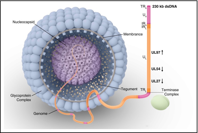
Figure 1.
Human CMV virion structure and structural components. The genome is packaged in an icosahedral capsid surrounded by a lipid envelope. Viral phosphoproteins (eg, pp65 antigen) are found in the tegument, a space between the nucleocapsid and the envelope. The DNA genome is formed by 2 covalently linked genome segments (L, long; S, short), each consisting of a central unique region (UL, unique long; US, unique short). These unique regions are flanked by inverted repeats either at the ends (IR, inverted repeat; TR, terminal repeat) or internally at the intersection of the long and short segments (IRL, internal repeat long; IRS, internal repeat short). Vertical arrows show the direction of the open reading frame for each gene of interest for drug resistance (UL97, UL54, and UL27). During CMV replication, a single long DNA chain is synthesized. This chain contains multiple repeated gene sequences known as concatemers. Each concatemer is cleaved into multiple gene sequences known as monomers, forming the genetic material for each virion. This process of replication, cleavage, and packaging is performed by a terminase complex. dsDNA, double-stranded DNA.

