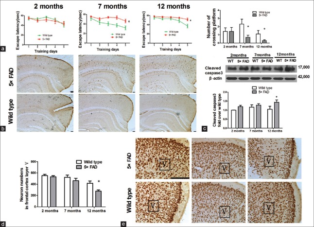
Figure 1.
The declined cognition in 7-month-old 5×FAD mice and the loss of neurons in the frontal cortex of the 12-month-old ones. (a) Morris water maze test. Escape latency and the number of crossings over the platform in 7- and 12-month old 5×FAD mice (n = 10–14, *P < 0.05, †P < 0.01 vs. age-matched wild-type mice). (b) 6E10 staining in 2-, 7-, and 12-month-old 5×FAD mice and wild-type mice. Scale bar = 100 μm. (c) Western blots analysis showed that activated caspase-3 increased in 5×FAD mice at 12 months of age (n = 8, *P < 0.05 vs. wild-type mice). (d) The neurons within the fifth layer of the cortex was quantified (n = 6, *P < 0.05 vs. wild-type mice). (e) NeuN staining for neural nuclei in the frontal cortical slices from 2-, 7-, and 12-month-old mice. Scale bar = 100 μm; 5×FAD: Transgenic mice with five familiar Alzheimer's disease; WT: Wild-type mice; Aβ: Amyloid β; V: The fifth layer of the frontal cortex.

