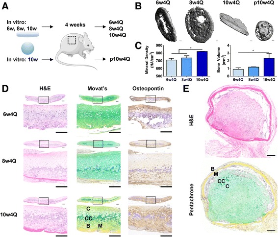
Fig. 6.

Evaluation of implantation outcomes by histological and μCT analysis. a Discs cultured for 6 weeks, 8 weeks or 10 weeks, as well as pellets cultured for 10 weeks were implanted subcutaneously in mice. After 4 weeks, tissues were explanted for analysis. b Representative μCT images show that mineralization of discs increased with length of culture, and was associated with an early mineralization at the rim. Whereas the discs formed a sheet of mineral, the pellet formed a spherical shell. c Mineral density and volume increased with the length of culture; 10w4Q discs exhibited higher mineral density and volume than 6w4Q and 8w4Q discs. d Whole tissue and detailed histological stains of discs after endochondral ossification. Scale bar: 200 μm. Discs were more mineralized with length of culture and mature bone was seen in 10w4Q discs. Bone formed at the bottom of the discs and the infiltrating marrow-like cells were observed. Non-calcified cartilage remained at the surface. e Whole tissue histological stains of pellets after endochondral ossification. Scale bar: 200 μm. Bone formed at the surface, with infiltrating marrow-like cells beneath gradually resorbing the calcified cartilage. H&E hematoxylin and eosin
