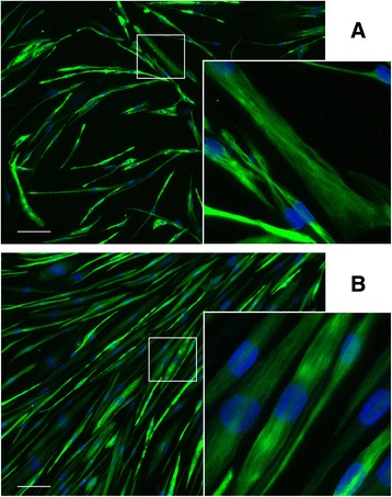
Fig. 1.

Representative images of primary and immortalized human myotubes. Myogenic cells were purified by magnetic bead sorting of CD56 expression then differentiated for 5 days. Examples here are from primary (a) and immortalized (b) populations of a healthy subject (CHQ). Myotubes were immunostained for desmin (green) to determine the percentage of myogenic purity (>94% for all samples). Magnified region shows the structure of desmin filaments in both primary and immortalized cells. Nuclei are DAPI-stained (blue). Scale = 100 μm
