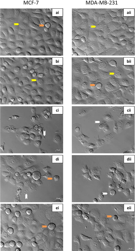
Fig. 3.

PlasDIC images of MCF-7 and MDA-MB-231 cells exposed to the compound with/or without 3MA for 24 h. i MCF-7 cells and ii MDA-MB-231 cells grown in a DMSO and b 3MA served as negative controls. Confluent cell growth with no signs of cell distress was demonstrated. c Actinomycin D (0.1 μg/ml) served as a positive control for apoptosis, resulting in apoptotic body formation and compromised cell density. d ESE-15-ol-treated cells revealed the presence of rounded cells, formation of apoptotic bodies and decreased cell density. e ESE-15-ol exposure to cells in which autophagy had been inhibited with 3MA showed an increase in cell viability. (Arrow colour key: Yellow = interphase cells; orange = rounded cells in metaphase; white = apoptotic bodies.)
