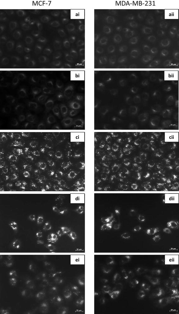
Fig. 4.

Fluorescent microscopy with monodansylcadaverine staining of MCF-7 and MDA-MB-231 cells. i MCF-7 and ii MDA-MB-231 cells treated with a DMSO and b 3MA served as negative controls and displayed non-specific MDC staining. c Tamoxifen was used as a positive control for acidic vacuoles and displayed clear MDC stained vacuoles. d ESE-15-ol treated cells showed increased MDC staining, while e ESE-15-ol treated cells together with 3MA showed less distinctive MDC staining, indicating partial autophagy inhibition (40× magnification)
