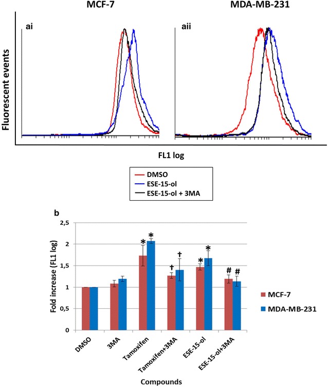
Fig. 9.

LC3 fluorescence determination within MCF-7 and MDA-MB-231 cells after a 24 h exposure. Overlay histogram of ai MCF-7 and aii MDA-MB-231 exposed cells. Cells treated with ESE-15-ol showed a right shift which was greater than ESE-15-ol with 3MA. b Graphical representation shows an increase in LC3 detection within ESE-15-ol-treated cells. Bars represent averages of three biological repeats (P value <0.05; standard deviations represented by T-bars; *indicates statistical differences between compounds and DMSO vehicle control; #indicates statistical difference between ESE-15-ol and ESE-15-ol with 3MA; †indicates statistical difference between tamoxifen and tamoxifen with 3MA)
