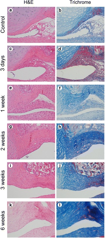
Fig. 3.

Serial microscopic findings of axillary recess of the glenohumeral joint in the control and immobilization groups. a, b Microscopic findings show no inflammation or fibrosis in the control group (H&E and Trichrome, ×200). c, d Inflammation with mild fibrosis is identified at 3 days after immobilization (H&E and Trichrome, ×200). e, f Fibrosis with mild inflammation is identified at 1 week after immobilization (H&E and Trichrome, ×200). g, h Fibrosis with minimal inflammation is identified at 2 weeks after immobilization (H&E and Trichrome, ×200). i, j Fibrosis with minute inflammation is identified at 3 weeks after immobilization (H&E and Trichrome, ×200). k, l Fibrosis without inflammation is identified at 6 weeks after immobilization (H&E and Trichrome, ×200)
