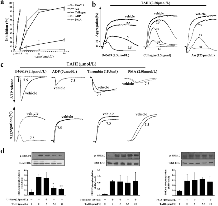
Figure 2. TAIII selectively inhibits TxA2-mediated platelet activation.
Rat PRP was preincubated with increasing concentrations of TAIII (0–60 μmol/L) or vehicle for 3 min and then stimulated with U46619 (2.5 μmol/L), AA (125 μmol/L), collagen (1 μg/ml), ADP (5 μmol/L) or PMA (250 nmol/L). (a) Dose-inhibition curves for TAIII were obtained from the results of three independent experiments, and data are presented as the percentage inhibition (mean ± SEM). (b) Typical platelet aggregation traces are representative of three independent experiments. (c) Rat PRP was preincubated with 7.5 μmol/L TAIII for 3 min and then stimulated with U46619 (2.5 μmol/L), ADP (5 μmol/L), and PMA (250 nmol/L). Incubation of washed platelets with TAIII (7.5 μmol/L) was followed by stimulation with thrombin (1 IU/ml). Platelet aggregation and ATP release were measured by a turbidimetric method. Typical real-time ATP secretion and platelet aggregation traces are representative of three independent experiments. (d) TAIII-treated platelets were stimulated with 2.5 μmol/L U46619, 1 IU/ml thrombin or 250 nmol/L PMA to induce ERK1/2 phosphorylation. Densitometric analyses of the blots were performed using Image J software. The relative protein expression levels are presented as the ratio of phosphorylated ERK1/2 levels to the corresponding total ERK1/2 levels. Images are representative of three independent experiments, and the results are presented as the mean ± SEM. Statistical significance was determined by Student’s t-test. *p < 0.05 and **p < 0.01, compared with U46619-treated platelets.

