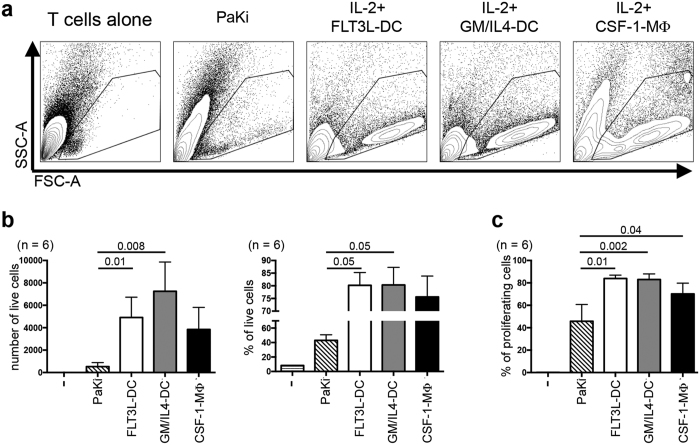
Figure 2. Characterization of T-cell allostimulatory capacity of P. alecto BM-derived DC and macrophages.
(a–c) Following 6 days of culture of BM mononuclear cells as described in Fig. 1, a mixed lymphocyte reaction (MLR) was carried out by co-culturing these differentiated cells with CFSE-labelled allogenic P. alecto bat lung (n = 4) or spleen (n = 2) mononuclear cells in the presence of recombinant human IL-2 (50 UI/ml) for another 6 days until Day 12 (D12). As controls, the lung or spleen cells were cultured 6 days alone (far left panel) or in the presence of PaKiT03 cells (PaKi, left panel). At D12 (6 days of MLR), cells were analysed by flow cytometry (see Supplementary Fig. 2a for the gating strategy) and (a) their size (FSC-A) and granulosity (SSC-A) were analysed. (b) The number (left panel) and proportion (right panel) of live cells in each condition described in panel (a) are displayed. (c) Proportion of proliferating cells (CFSElo, see Supplementary Fig 2 for CFSE histograms) among live cells in each condition described in panel (a) are displayed. P values were calculated using the paired t-test.

