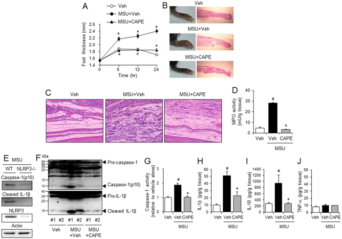
Figure 3. Oral administration of CAPE prevents MSU crystals-induced gout in mouse foot by blocking NLRP3 inflammasome activation.
Mice were orally administered CAPE (30 mg/kg) or vehicle (Veh, 0.02% DMSO in water). After 1 hr, MSU crystals (2 mg/0.1 ml of PBS/mouse) or PBS alone were subcutaneously injected into the pad of the right hind foot of each mouse. After 24 hr, the footpad tissue was collected for analysis. (A) Time course of foot thickness. (B) Representative photographs and H&E staining of the hind feet. (C) Infiltrated neutrophils in the hind foot tissue appear as purple dots in H&E staining (400X). (D) Supernatants from the foot tissue lysates were analyzed for myeloperoxidase (MPO) activity. (E) MSU crystals (2 mg/0.1 ml of PBS/mouse) or PBS alone were subcutaneously injected into the pads of the right hind feet of wild-type (WT) and NLRP3-knockout mice. The foot tissue was analyzed by immunoblotting for caspase-1(p10), IL-1β, NLRP3, and actin. (F–J) The foot tissues from Fig. 3A were subjected to immunoblotting for pro-caspase-1, caspase-1(p10), pro-IL-1β, and IL-1β, a caspase-1 enzyme activity assay, and ELISAs for IL-1β, IL-18, and TNF-α. The values in the line and bar graphs represent the means ± SEM (n = 3 mice). #Significantly different from vehicle alone, p < 0.05. *Significantly different from MSU alone, p < 0.05. Veh, vehicle.

