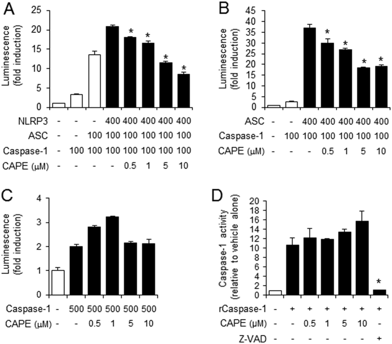
Figure 4. The suppression of inflammasome activation by CAPE is dependent on ASC.
(A–C) 293T cells were transiently transfected with the iGLuc luciferase reporter plasmid (100 ng) and expression plasmids. Luminescence derived from iGLuc activation in each sample was normalized by β-galactosidase activity transfected as an internal control in each sample. (A) *Significantly different from NLRP3 + ASC + caspase-1, 0.5: p = 0.0064, 1–10: p =< 0.0001. (B) *Significantly different from ASC + caspase-1, 0.5: p = 0.0036, 1: p = 0.0001, 5–10: p =< 0.0001. (D) In vitro assay for caspase-1 enzyme activity was performed using a fluorometric caspase-1 assay kit with recombinant human caspase-1 (rCaspase-1; Bio-vision) in the presence or absence of CAPE or Z-VAD-FMK according to the manufacture’s instruction. The fluorescence was recorded at 400 nm after excitation at 505 nm with SpectraMaxM5 (Molecular Devices, Sunnyvale, CA). *Significantly different from rCaspase-1 alone, p = 0.0007. The values represent the means ± SEM (n = 3).

