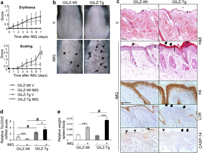
Figure 2. GILZ-Tg mice show increased psoriatic features in response to imiquimod (IMQ).
(a) Score of the erythema and scaling in GILZ-Wt and GILZ-Tg for the duration of the experiment. V: vehicle; IMQ, imiquimod. (b) Macroscopical appearance of GILZ-Wt and GILZ-Tg mice treated with IMQ at day 7. (c) Representative Hematoxilin&Eosin (H&E) stained and immunohistochemistry sections of adult mouse skin of IMQ-treated GILZ-Wt and GILZ-Tg mice. Epidermal thickening (brackets), hyperkeratosis (asterisks), abnormal differentiation with parakeratosis (thick arrows), and epidermal infiltrate (Munro-like abscess, arrowhead) are indicated. Immunostaining with keratin (K)6, Loricrin (LOR), and Caspase-14 (CASP-14) antibodies is also shown. Note the increased dermal cellularity in IMQ-treated GILZ-Tg relative to GILZ-Wt mice. Bar: 50 μm. Samples from two independent experiments were assessed (n = 15 GILZ-Wt, n = 16 GILZ-Tg). (d) Relative Tsc22d3/Gilz mRNA levels in GILZ-Wt and GILZ-Tg mouse skin were assessed by RT-QPCR before and after treatment with IMQ at day 7. (e) Splenomegaly was determined by the weight ratio of spleen relative to body. (d,e) Mean values ± SD are shown. Post hoc Tukey test #p < 0.05; ***p < 0.001, n = at least 5 per genotype and treatment. Asterisks: significance between treatments within each genotype; hashes: significance between genotypes in the same treatment group.

