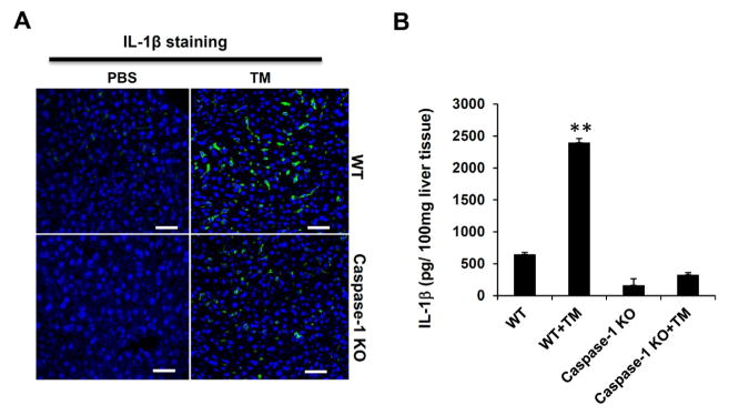
Figure 4.
ER stress-induced hepatic steatosis is associated with inflammasome activation and IL-1β production. (A) IL-1β immunostaining of liver sections. IL-1β levels in liver tissues from WT and caspase-1KO mice treated with or without TM were detected by immunostaining with an anti-IL-1β specific antibody. The green color indicates IL-1β, and nuclei are stained in blue by DAPI. Scale bar: 100 μm. (B) Liver tissues from WT and caspase-1 KO mice treated with TM treatment were homogenized, and analyzed by ELISA. Data are presented as means ± SEM. **P<0.01 (Student’s t test).

