Figure 1.
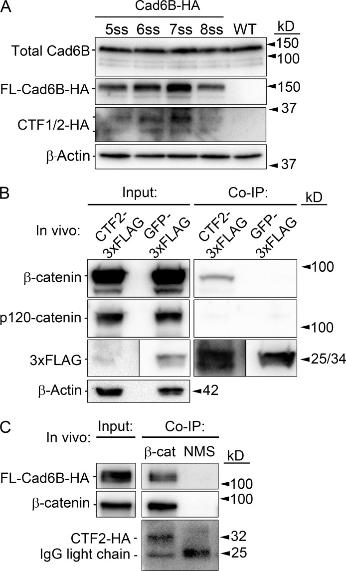
Cad6B CTF2s are generated before and throughout cranial neural crest EMT and remain associated with β-catenin. (A) Cad6B is subjected to γ-secretase–mediated proteolysis before, and during, EMT. Premigratory neural crest cells were electroporated at 2–3ss, and cranial neural folds were collected at specific stages for HA immunoblot analysis. (B) CTF2 physically associates with β-catenin, but not p120-catenin, in vivo. Embryo midbrains were electroporated at 4ss and incubated beyond the initiation of EMT. Dorsal neural tubes containing CTF2-3xFLAG– or GFP-3xFLAG–expressing neural crest cells were subjected to coimmunoprecipitation (co-IP) and immunoblotting. (C) HA-tagged CTF2s created via full-length Cad6B-HA proteolysis remain bound to β-catenin in cranial neural crest cells in vivo. Embryos were dorsal/ventrally electroporated at 4ss, and dorsal neural tubes were collected at 8ss. Normal mouse serum (NMS) immunoprecipitation with Cad6B-HA lysate served as a negative control. WT, wild type.

