Figure 5.
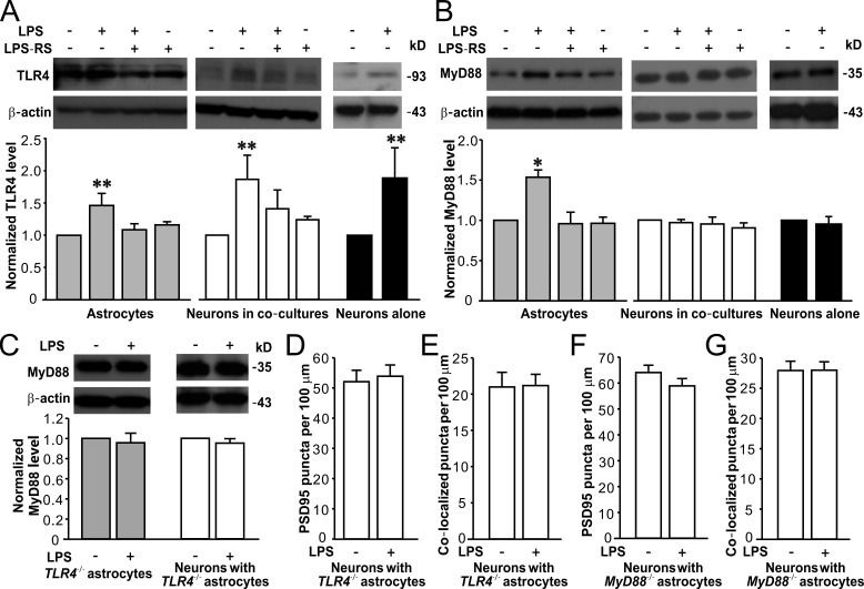
LPS specifically activates TLR4–MyD88 signaling in astrocytes. Representative Western blots of TLR4 (A) and MyD88 (B) in astrocytes, neurons in co-cultures (DIV15), and neurons alone (DIV15; top) and quantification of TLR4 (A) and MyD88 (B) expression (bottom). n = 4, 4, and 3 for astrocytes, neurons in co-cultures, and neurons alone groups, respectively. (C) Representative Western blots of MyD88 in TLR4−/− astrocytes and neurons co-cultured with TLR4−/− astrocytes (top) and quantification of MyD88 expression (bottom). n = 3 for each group. Quantification of PSD95 puncta (D) and PSD95/synaptophysin colocalized puncta (E) in neurons co-cultured with TLR4−/− astrocytes. n = 32 in each group. Quantification of PSD95 puncta (F) and PSD95/synaptophysin colocalized puncta (G) in neurons co-cultured with MyD88−/− astrocytes. n = 29 for each group. β-Actin is shown as a loading control. *, P < 0.05; **, P < 0.01 (t test or one-way ANOVA with the post hoc Dunnett’s test). Data are mean ± SEM.

