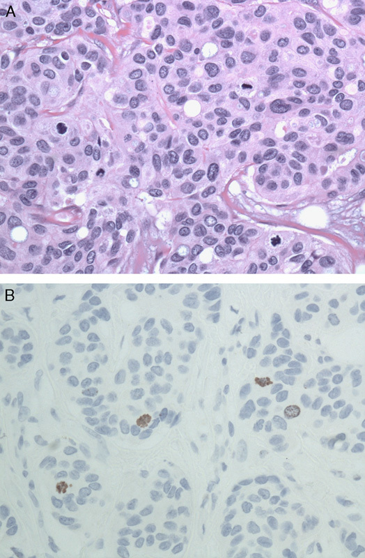
FIGURE 2.

A, It may be difficult to determine if a potential mitotic figure on hematoxylin and eosin is truly a mitotic figure (×400). B, Immunohistochemistry stain for PhH3 highlights mitotic figures (×400). PhH3 indicates phosphorylated histone H3.
