Figure 1.
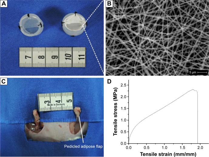
The rat chamber model and microstructure, stress–strain curve of PCL nanofibrous mesh.
Notes: (A) Cylindrical silicone chambers (left), PCL nanofibrous mesh was attached to the internal surface of the chamber (right). (B) SEM images of PCL nanofibrous mesh demonstrated the pore structure and nano-sized fibers. (C) A vascularized pedicled adipose flap based on the superficial inferior epigastric vessels of standardized dimensions was dissected in situ. (D) The stress–strain curve of PCL nanofibrous mesh.
Abbreviations: PCL, polycaprolactone; SEM, scanning electron microscopy.

