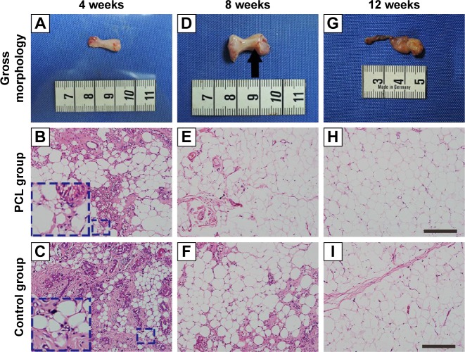
Figure 3.
Engineered adipose flap in the PCL group chamber and hematoxylin and eosin-stained section at different time points.
Notes: The tissue of PCL group from week 4 consisted largely of a fragile, white, gel-like fibrin exudation surrounding the flap (A) with small adipocytes (inset) detectable in the connective tissue (B). By 8 weeks, new adipose tissue had formed (E) and small blood vessels could be detected on the surface of the fibrin exudation (D). Similar observations were made ain 4- and 8-week constructs of control groups (C, F), By week 12, both groups had developed vascularized and well-organized adipose tissue, with almost nonexistent fibrin exudation at the outermost layer (G, H, I). Scale bar =100 μm.
Abbreviation: PCL, polycaprolactone.

