
Figure 5.
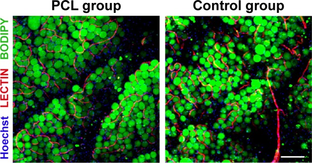
Visualization of engineered adipose tissue at week 12 by whole-mount staining.
Notes: Interaction of blood vessels and adipocytes in engineered adipose tissue (PCL group) was demonstrated by Lectin/BODIPY/Hoechst (red/green/blue) staining and compared with adipose tissue in control group. At baseline, capillaries or vessels ran between mature adipocytes in both groups, and adipose tissue in the PCL group displayed a more mature and integrated structure than the control group. Scale bar =200 μm.
Abbreviation: PCL, polycaprolactone.