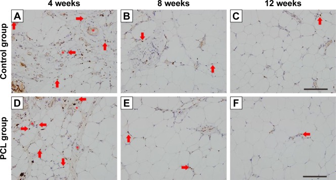
Figure 6.
Ki67-stained section of the samples at different time points.
Notes: In both groups, most Ki67-positive cells (arrowhead) were located near blood vessels (star) at week 4 (A, D) and the number decreased at week 8 (B, E). Almost no Ki67-positive cells could be found at week 12 (C, F). Scale bar =200 μm.
Abbreviation: PCL, polycaprolactone.

