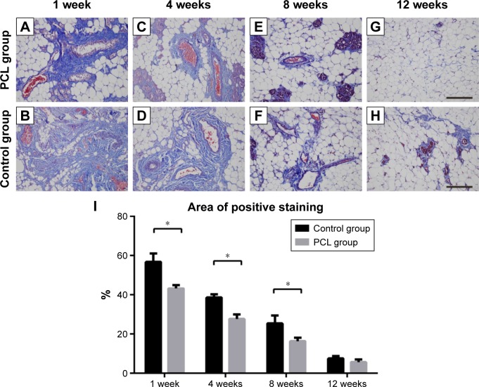
Figure 7.
Collagen deposition at different times.
Notes: From weeks 1 to 4, massive collagen deposition, limited to the areas close to the pedicle, were found in both groups (A–D), then the collagens were gradually replaced by newly formed small adipocytes by week 8 (E, F). Statistical analysis demonstrated that the control groups still had higher collagen content between weeks 1 and 8 (I). At week 12, there was a further decrease in the amount of collagens, and the remaining collagens were observed mainly near small blood vessels (D, H). All data were expressed as mean ± standard deviation. *P<0.05 compared between PCL group and control group. Results are in response to an analysis of Student’s t-test of two groups. Scale bar =200 μm.
Abbreviation: PCL, polycaprolactone.

